Molecular imaging leads to early detection of glaucoma
【PLoS One. 2012;7(1):e30526】
-
Glaucoma is a progressive disease leads to visual deficit, and early detection and therapy of glaucoma are crucial.
Our research team, in collaboration with Professor Hara's team at Gifu Pharmaceutical University, investigated what actually happens in the brains of glaucoma in an experimental glaucoma model monkey using PET.
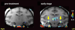 We focused on finding the inflammatory reaction, particularly, activated microglia, which is known as one of important immune cells in the brain. PET imaging in the glaucomatous monkey with its early-stage revealed that microglia-associated PET probes were accumulated in the deep in the brain, at the lateral geniculate body. This microglial activation was also accompanied by shrinkage and degeneration of neurons in the lateral geniculate body in a histopathological examination. These results suggest the possibility that glaucoma, which has been considered to be a disease of eye, could be correctly diagnosed by brain PET imaging at the initial stage of the disease. Glaucoma is a slowly progressive disease of eye, and is the leading cause of blindness. Early detection of glaucoma is crucial for preventing its progress and for reducing the blind.