


Technologies of CLST
PET imaging
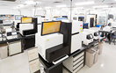
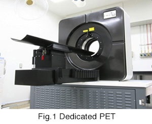 Two dedicated PET scanners (Fig.1), two combined PET/CT scanners (Fig. 2) and one CT scanner (Fig. 3) are installed in RIKEN CLST-DBDI Kobe. The all scanners have high spatial resolution for small animal study. PET image data is the 3-diemsional numerical data of distribution of radioisotopes. And the following kinds of image data can be obtained.
・Static image data
・Dynamic image data
・Whole body image data
・Dynamic whole body data
・Gated image data
Static image data is the simple 3-dimensinal RI distribution data. Dynamic image data is the time-course image data, which is the 4-dimentional RI distribution data. Whole body data is the 3-dimensional image data which coves longer range than scanner range by using bed motion. Dynamic whole body image is the time-course image of RI distribution in whole body, but time frame definition must be much longer than simple dynamic image. Gated image data is the RI distribution data synchronized to cyclic motion such as the beat of heart or respiration.
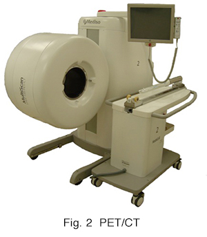
90-degree tilting of scanner is possible for PET/CT. By using the tilting, a primate animal scan with visual stimulation can be performed.
The field of view of PET and PET/CT scanners is cylindrical region, which is maximum 20 cm in diameter and 7.8 cm in axial for PET, and 20 cm in diameter and 15 cm in axial for PET/CT. These scanners can perform scan of animal from rodent such as mice and rats, to primate animal. And the whole body imaging is possible by using the scan with bed motion, which is called “Whole body scan”. Image data is saved into the image file as 3-dimentional matrix data format in the unit of “Bq/ml”, it is radio activity concentration. PET part of PET/CT and dedicated PET has high sensitivity and perform scan in 3-diemnsional acquisition mode which is not sorted into each slices at the data acquisition. Then, 3-dimensonal image reconstruction algorithm is used to get image data.
The PET acquired data is the coincidence detected gamma photon information includes time and two detectors position, and saved the sequential data stream called list-mode data set. The process of sorting into sinogram of dynamic or static is the post process. The time-course analysis of radiopharmaceutical such as pharmacokinetics (PK) or pharmacodynamics (PD) is able to be done by using dynamic image data. The PET/CT and PET can cover the mouse whole body by simple static or dynamic scan. For bigger animal than mouse, dynamic whole body scan can be used, which is the performing simple scan with bed motion repeatedly. If you input the gate signal to the scanner which is synchronized to heart beat or respiration, gated image can be obtained. The choice of gated or non-gated image is made after the finish the scan. It is the post processing as dynamic image.
PET and PET/CT have the function of 45, 90-degree tilting respectably. This function makes PET scan with pseudo-sitting or sitting scan performable. By using this, images of brain activation changes by visual stimulation can be obtained.
Measured data of PET is not image data. Image data is processed data of measured data. This process is called “Image reconstruction”. The image reconstruction is a kind of solving inverse problem. There are two major algorithm for image reconstruction. One is Filtered Backprojection (FBP) based on mathematical analysis, and standard algorithm for tomography. The other is Maximum Likelihood – Expectation Maximization (ML-EM) method based on the statistics. The solution of ML-EM is not strait forward equation, it is required iterative calculation. Then, this method is called “Iterative”. For FBP, accuracy of image data is high, but noise level is high. For iterative, image is clear (low noise), but low accuracy if the parameters in reconstruction process are not proper. In RIKEN CLST-DBDI Kobe, reconstruction parameters are fixed by considering the purpose of the study and accuracy of data to obtain image data as molecular imaging.
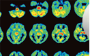
PET and CT images can be obtained by single PET/CT scanner, and fusion image of PET and CT can be got. CT image contains mainly anatomical information of subject, and PET image contains RI distribution information. Although, getting anatomical information is difficult by only PET scanner, it can be done by using CT image scanned by dedicated CT scanner also installed in RIKEN CLST-DBDI Kobe.
Attenuation correction is one of important corrections, affects the image data accuracy. There are several methods of attenuation correction data for dedicated PET. One is the transmission method with external point source. others are using subject shape information getting by PET image and combination of numerical model image data. Attenuation correction is selected by the situation of PET scan protocol and using animal holder.
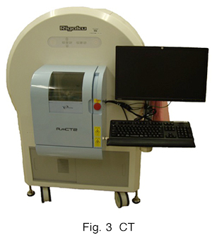
CT is the scanner to get attenuation distribution by exposing X-ray. CT is much familiar than PET in a hospital. A dedicated CT which has high spatial resolution for a rodent animal is installed in RIKEN CLST-DBDI Kobe. Image data is 3-dimentional matrix numerical data, and data unit is Haunsfiled Unit (HU). HU is the value of attenuation, defined air is -1000, water is zero. Bone vale is usually more than 400. Although, HU value is not linear attenuation correction value, it is commonly used in a hospital because of compensating the X-ray energy dependency and can be used for quantitative analysis.
Small animal CT is using advance technology called “Cone-beam CT (CBCT)”. The CBCT is also used in PET/CT for small animal too. CBCT acquires data in 3-dimensional similar to PET and needs 3-dimensional image reconstruction algorithm. Generally, the reconstruction algorithm used in CT is FBP. Radiation dose of CT, parameters in reconstruction process are selected by considering restriction of noise level in image data and size of the target region etc.
In RIKEN CLST-DBDI Kobe, PET, PET/CT and CT are operating to obtain image data as scientific data, not just clear image.