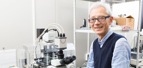
RIKEN Center for Life Science Technologies

Topics

  • Research

    Latest publications from CLST.
     
  • News

    Awards, agreements, and other news.
  • Events

    Upcoming and past events including seminars, symposiums, open days.

For Public

To let you know about our research, this area contains 4 types of information about CLST; “Article”, “Videos”, “Event” and “Study”.
At “Article”, you can read articles on interviews and lectures, and you can enjoy the videos about CLST at “Videos”. If you want to meet and talk directly with the researcher, “Visit” give you some information of such events. You can find more difficult contents to know about our research deeply at “Study”.
  • Article
  • Event
  • Videos
  • Study

Labs & Technologies

Ultrastructural Research Team

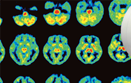
We obtain and analyze ultrastructural information of cells and tissues by utilizing electron microscopy for elucidation of principles of life towards applied research.


* Due to the reorganization starting as new centers in April 2018, this laboratory is now belong to the Center for Biosystems Dynamics Research. As for the latest information, please see the following URL below.
> The webpage of Laboratory for Ultrastructural Research, Center for Biosystems Dynamics Research

Team Leader
Shigenobu Yonemura  Ph.D.

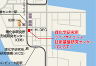
2-2-3 Minatojima-minamimachi, Chuo-ku, Kobe, Hyogo 650-0047, Japan
Tel: +81-78-306-3105

3_11_yonemura.png

>>> Lab Site

Research Area

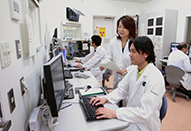
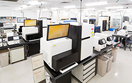
This research team analyzes detailed morphological information in various researches of the life science field, which is acquired through ultrastructural techniques using transmission and scanning electron microscopy. In collaboration with many laboratories including within RIKEN, we have accumulated a large amount of experiences to improve methods for better morphology.

Main Publications List

2

Maintenance of stereocilia and apical junctional complexes by Cdc42 in cochlear hair cells.

Ueyama T, Sakaguchi H, Nakamura T, Goto A, Morioka S, Shimizu A, Nakao K, Hishikawa Y, Ninoyu Y, Kassai H, Suetsugu S, Koji T, Fritzsch B, Yonemura S, Hisa Y, Matsuda M, Aiba A, Saito N.
J Cell Sci, 127(Pt9), 2040-2052 (2014).
3

Transplantation of embryonic and induced pluripotent stem cell-derived 3D retinal sheets into retinal degenerative mice.

Assawachananont J, Mandai M, Okamoto S, Yamada C, Eiraku M, Yonemura S, Sasai Y, Takahashi M.
Stem Cell Reports, 2(5), 662-674 (2014).
4

Sphingomyelin clustering is essential for the formation of microvilli.

Ikenouchi J, Hirata M, Yonemura S, Umeda M.
J Cell Sci, 126(Pt16), 3585-3592 (2013).
5

NMII Forms a Contractile Transcellular Sarcomeric Network to Regulate Apical Cell Junctions and Tissue Geometry.

Ebrahim S, Fujita T, Millis BA, Kozin E, Ma X, Kawamoto S, Baird MA, Davidson M, Yonemura S, Hisa Y, Conti MA, Adelstein RS, Sakaguchi H, Kachar B.
Curr Biol, 23(8), 731-736 (2013).
6

Talin couples the actomyosin cortex to the plasma membrane during rear retraction and cytokinesis.

Tsujioka M, Yumura S, Inouye K, Patel H, Ueda M, Yonemura S.
Proc Natl Acad Sci U S A, 109(32), 12992-12997 (2012).
7

Self-formation of functional adenohypophysis in three-dimensional culture.

Suga H, Kadoshima T, Minaguchi M, Ohgushi M, Soen M, Nakano T, Takata N, Wataya T, Muguruma K, Miyoshi H, Yonemura S, Oiso Y, Sasai Y.
Nature, 480(7375), 57-62 (2011).
9

Cadherin-actin interactions at adherens junctions.

Yonemura S.
Curr Opin Cell Biol, 23(5), 525-522 (2011).
10

alpha-Catenin as a tension transducer that induces adherens junction development.

Yonemura S, Wada Y, Watanabe T, Nagafuchi A, Shibata M.
Nat Cell Biol, 12(6), 533-542 (2010).

Member  *concurrent

CLST was reorganized into three centers according to the RIKEN 4th Medium-Term Plan from April 1, 2018. For the latest information of Ultrastructural Research Team, please visit the following websites.


> The webpage of Laboratory for Ultrastructural Research, Center for Biosystems Dynamics Research [http://www.bdr.riken.jp/en/research/labs/yonemura-s/index.html]